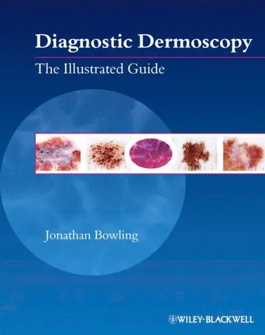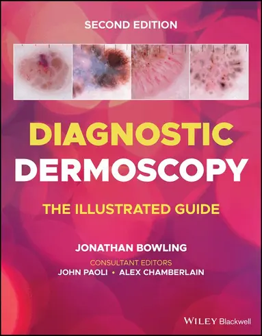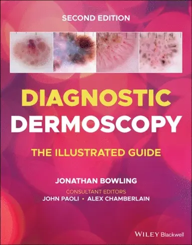
Diagnostic Dermoscopy
The Illustrated Guide
E-bok
Engelska, 2011844 kr
Tillfälligt slut. Bevaka för att få ett mejl när produkten kan beställas.
Fler format och utgåvor
Beskrivning
Ideal for clinic use, both for diagnosis and for explaining to the patient, this guide provides: * A quick reference atlas guide to the diagnosis skin lesions, especially, but not limited to, those that are cancerous * Icons for each condition linked to high definition dermoscopy and clinical photographs * Real dermatoscopic images and the associated clinical photographs on the page opposite
Produktinformation
- Utgivningsdatum:2011-11-01
- Språk:Engelska
- ISBN:9781444329834
- Förlag:John Wiley and Sons Ltd
- Dimensioner:150 x 250 x 15 mm
Utforska kategorier
Mer om författaren
Jonathan Bowling, MD, MRCP is consultant dermatologist and honorary senior clinical lecturer at the University of Oxford Department of Dermatology. He has been providing training in dermoscopy since 2002. To date over 500 UK doctors have attended his national one day courses in dermoscopy education. He is?a board member of the International Dermoscopy Society and lectures?internationally on dermoscopy.
Innehållsförteckning
- Introduction to dermoscopy 1. Introduction2. Instrumentsa. Introductionb. Non-polarised devicesc. Polarised devicesd. Hybrid devicese. Maintenance tips3. Normal Skina. Face, body and phototypeb. Photodamage4. Pigment depth and colour5. Dermoscopic alphabeta. Global featuresb. Local featuresMelanocytic lesions6. Melanocytic criteriaa. Pigmented networkb. Pigmented globulesc. Homogeneous pigmentationd. Parallel patterne. Cobblestonef. Starburst pattern7. Pigmented network8. Atypical pigmented network9. Cobblestone10. Globular11. Dermal naevi12. Blue naevi13. Halo naevi14. Recurrent naevi15. combined naevi16. Spitz naevia. Pinkb. Pigmented17. Naevi and phototypea. Skin phototype Ib. Skin phototype III18. Naevi & age19. Examples of predominant naevus morphologya. Globular homogeneousb. Patchy reticularc. Reticulard. Homogeneous20. Multicomponent21. Morphology22. Pigment distributionMelanoma23. 7 Features of melanomaa. Atypical pigmented networkb. Blue white veilc. Atypical blood vesselsd. Irregular dots & globulese. Irregular pigmentationf. Irregular streaksg. Regression structures24. Melanoma features and thicknessa. In-situb. Thin invasivec. Intermediate thickness invasived. Thick invasive25. Melanoma coloursa. Brownb. Black / hyperpigmentedc. Pink / hypopigmentedd. Multicoloured26. Melanoma typea. Superficial spreadingb. Nodulari. Pigmentedii. hypomelanoticc. Featurelessd. Smalle. Eccentricf. Cutaneousg. Negativeh. regression27. Algorithmsa. 7-point checklistb. ABCDc. Menzies scoring methodd. CASHe. 3-point checklistf. LimitationsNon-Melanocytic lesions28. Seborrhoeic keratosisa. Criteriab. Milia like cystsc. Comedo-like openingsd. Cerebriforme. Fingerprint structures29. Benign lichenoid keratosis30. Ink spot lentigo31. Solar lentigo32. Clear cell acanthoma33. Comedones34. Porokeratosis35. Haemangioma36. Lymphangioma37. Subcorneal haematoma38. Sebaceous gland hyperplasia39. Dermatofibromaa. DF vs Naevusb. Typical DFc. Non-typical DF40. Actinic keratosis41. Bowens disease42. SCC & Keratoacanthomas43. BCCa. Structuresb. Superficial BCCc. Nodular BCCd. Morphoeic BCCe. Pigmented BCCf. Brown BCC44. Blood vessels in skin lesions45. External pigmentation46. Radiotherapy scarsSpecial sites47. Acral lesionsa. Acral anatomyb. Acral naevic. Acral melanoma48. Facial lesionsa. Lentigo malignab. Lentigo maligna melanoma49. Nailsa. Melanonychiab. Nail unit melanomac. Nail unit SCCd. Erythronychiae. Subungual haematomaf. Nail unit Infectionsi. Bacterialii. Fungaliii. Viral50. Scalp tumoursa. Melanomab. BCCc. Naevus sebaceousd. Seborrhoeic keratosis51. Haira. Alopeciai. Non-scarring1. Androgenetic alopecia2. Alopecia areata3. Trichotillomaniaii. Scarring1. Lymphocytic scarring alopecia2. Neutrophlic scarring alopecia3. End stage scarring - pseudopeladeb. Miscellaneous hair conditionsi. Trichostasis spinulosaii. Ingrowing hairsiii. Pseudonits52. Mucosal lesionsa. Mucosal melanosisGeneral dermatology53. Skin infectiona. Scabiesb. Molluscum contagiosumc. Head liced. Viral warts54. Inflammatory skin diseasea. Psoriasis / eczemab. Lichen planusc. Blood vessel inflammation /Capillaritis vs vasculitisd. Urticaria pigmentosae. Granulomatous inflammationf. Dermatomyositis & sclerodermag. XanthogranulomaCase scenarios55. The nail lesion56. The foot lesion57. The hyperpigmented truncal lesion58. The atypical mole59. The purple ear lesionReference points60. 10 tips for better imaging61. 10 tips not to miss melanoma62. Key points for safe dermoscopy63. Skin lesion management pathwayImage gallerya. Naevib. Seborrhoeic keratosisc. Hypopigmented seborrhoeic keratosisd. BCC – nodulare. BCC – superficialf. BCC - pigmentedg. SCCh. Dermatofibromai. Haemangiomaj. Angiokeratomak. Melanoma – pinkl. Melanoma - brownm. Melanoma – blackn. Melanoma – arising in a naevuso. Melanoma – atypical vesselsp. Melanoma – multicolouredq. Melanoma - globulesr. Melanoma – regressions. Melanoma - extensive regressiont. Melanoma – blue white veil and atypical networku. Melanoma – SSMMv. Melanoma - blue white veilw. Melanoma – streaks
Hoppa över listan


Du kanske också är intresserad av
- Signerad!
SIGNERAD - Woka ur! Handbok i sydost- och östasiatisk matlagning
Vendela Wikström
Kartonnage, 2026
259 kr
- Signerad!
SIGNERAD - Mackans kost : Middagar och matlådor
Marcus Frank
Inbunden, 2026
4,8 utav 5 stjärnor. Totalt antal röster:(1094)
269 kr
- Signerad!